PETAlyze Open PETAlyze

PET/CT quantification in your browser

Open a PET/CT study, segment every uptake with a published threshold method, decide what is disease, and read total MTV, TLG and the Deauville score as you go. The images never leave your computer.

For research use only. Not for reporting patient examinations. Nothing to install: it runs in a recent desktop browser.

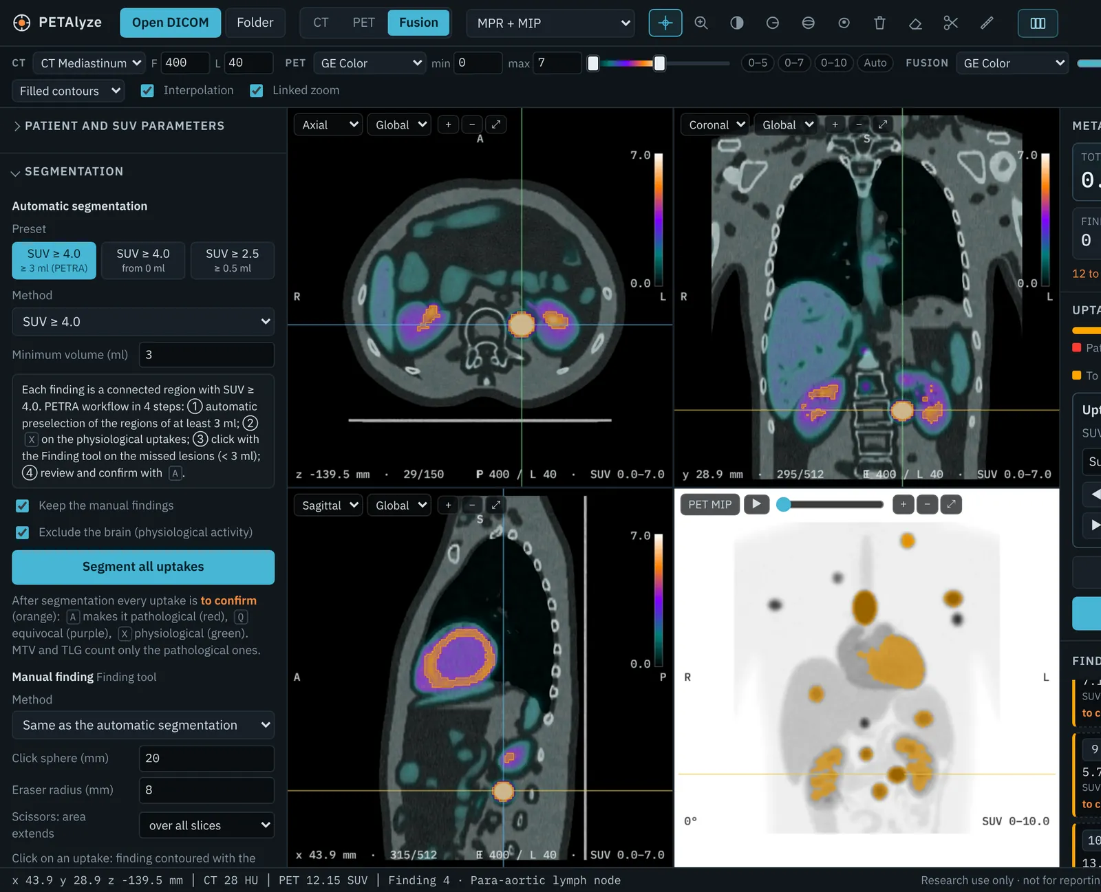
PETAlyze with a simulated lymphoma PET/CT: axial, coronal and sagittal fused views through a bulky mediastinal node, a MIP with pathological findings in red and physiological uptake in green, and the total MTV and TLG in the right panel.
The PET scale in GE Color, with the SUV 2.5 and 4.0 thresholds marked. Simulated lymphoma PET/CT.

How a study goes through PETAlyze

Six steps, from the DICOM folder to the report. Every number comes from a method you chose and a decision you confirmed. The images show a simulated lymphoma PET/CT, not a patient.

Open the study

Choose a folder, or drop DICOM files and ZIP archives onto the window. PETAlyze finds the CT and PET series, builds the volumes and converts PET to SUV from the DICOM header: body weight, lean body mass (SUL) or body surface area.

  • CT, PET and fused images in axial, coronal and sagittal planes, with a rotating MIP
  • GE Color, Hot Iron, Rainbow and other scales, set separately for PET and fusion
  • CT window presets and a two-handle slider for the PET scale
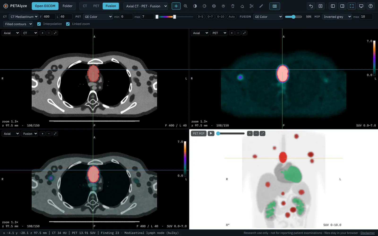
Axial CT, PET and fused PET/CT side by side through a bulky mediastinal lymph node, with the PET MIP.
Axial CT, PET and fusion side by side.

Segment every uptake

One click segments the whole body with the method you choose. With a fixed threshold, every voxel of a finding is above the threshold. Per-lesion methods contour each uptake from its own maximum.

SUV ≥ 4.0
PETRA preselection of regions of at least 3 ml, or of any volume
SUV ≥ 2.5
Fixed threshold
41% of SUVmax
Isocontour of each lesion
PERCIST
1.5 × liver SUVmean + 2 SD
A50P
50% of the background-corrected SUVpeak
MV2 and MV3
Majority vote of SUV 2.5, SUV 4.0, 41% and A50P
Extended MTV
SUV ≥ 4.0, plus A50P for lesions below 4

The brain can be marked as physiological automatically.

Segmentation panel with the SUV 4.0 PETRA preset selected, and every uptake contoured in orange, still to be confirmed.
After segmentation every uptake is orange: still to be confirmed.

Confirm each uptake

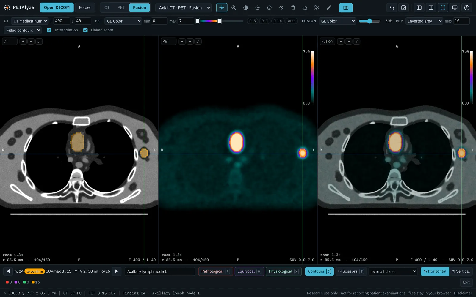
The review view puts axial CT, PET and fusion side by side, without side panels. Step through the uptakes and classify each one from the keyboard.

  • To confirm
  • Pathological A
  • Equivocal Q
  • Physiological X

When physiological activity touches a lesion, the scissors cut it away as a separate finding. The site of each uptake is proposed from the CT: lymph node stations, bone and vertebral level, liver, spleen, lung lobe.

Review view with axial CT, PET and fusion of an axillary lymph node, and the bar to classify it as pathological, equivocal or physiological.
Review view: one uptake at a time, with its site.

Measure

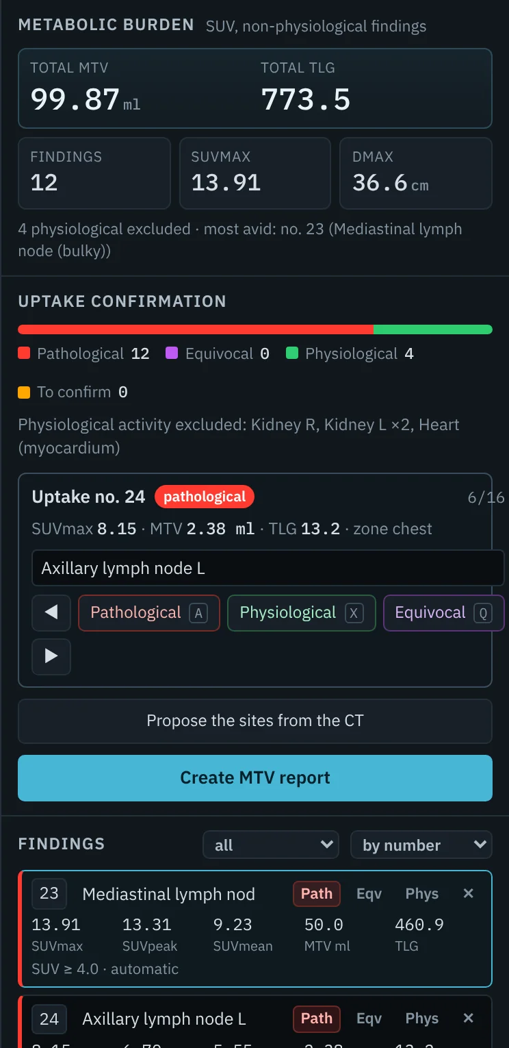
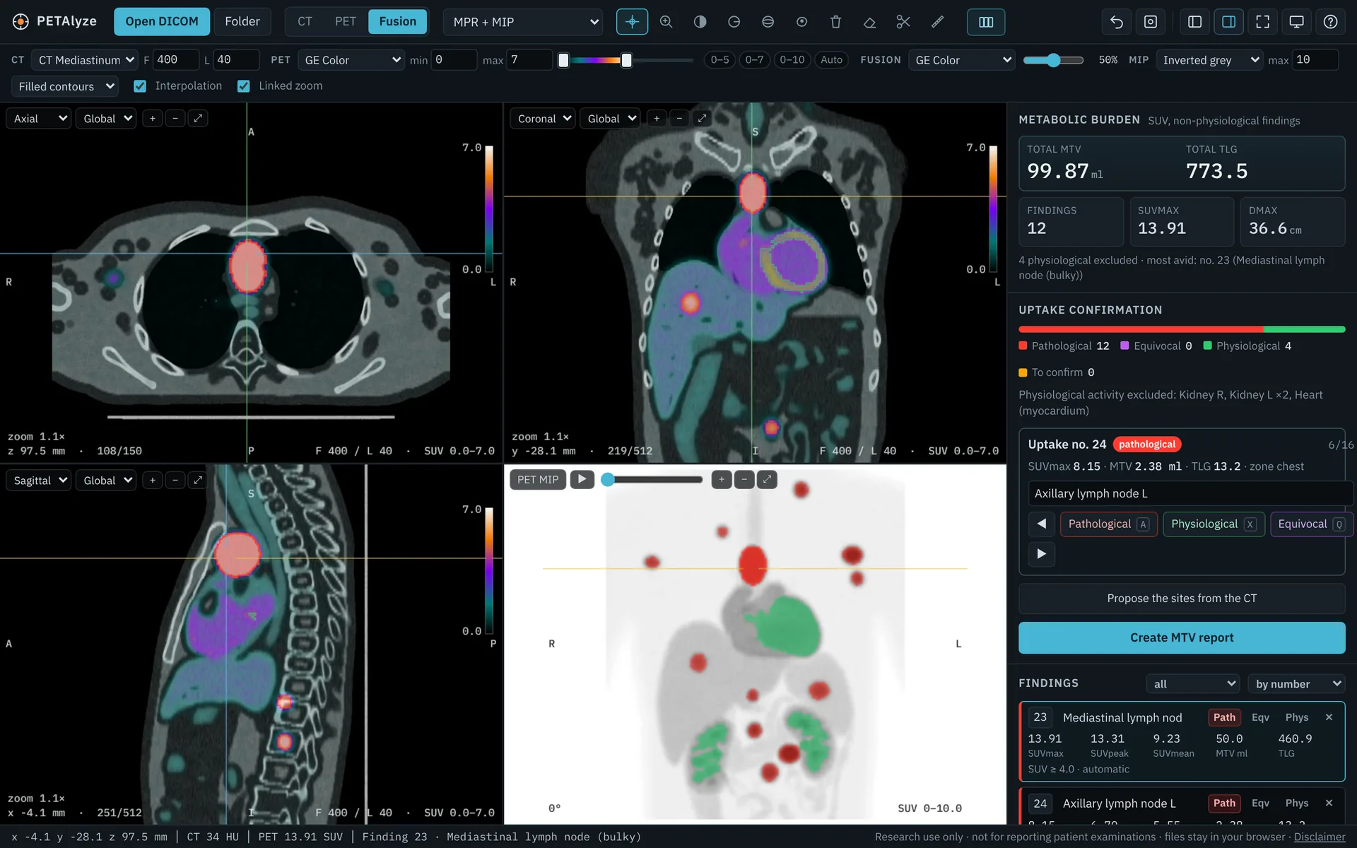
Total MTV, TLG and Dmax update with every decision, and count only the findings you called pathological. Each lesion shows SUVmax, SUVpeak, SUVmean, MTV and TLG, and you can compare all the methods on the same lesion.

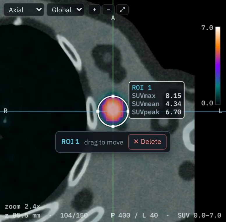
ROIs and VOIs show SUVmax, SUVmean and SUVpeak beside them. Click one to move it or delete it.

Results panel: total MTV 99.87 ml, total TLG 773.5, twelve findings, SUVmax 13.91, Dmax 36.6 cm, and the uptake confirmation counts.
Metabolic burden of the confirmed lesions.
A circular ROI on an axillary node with SUVmax 8.15, SUVmean 4.34 and SUVpeak 6.70 beside it, and the bar to move or delete it.
An ROI with its values, selected to move or delete.

Score Deauville

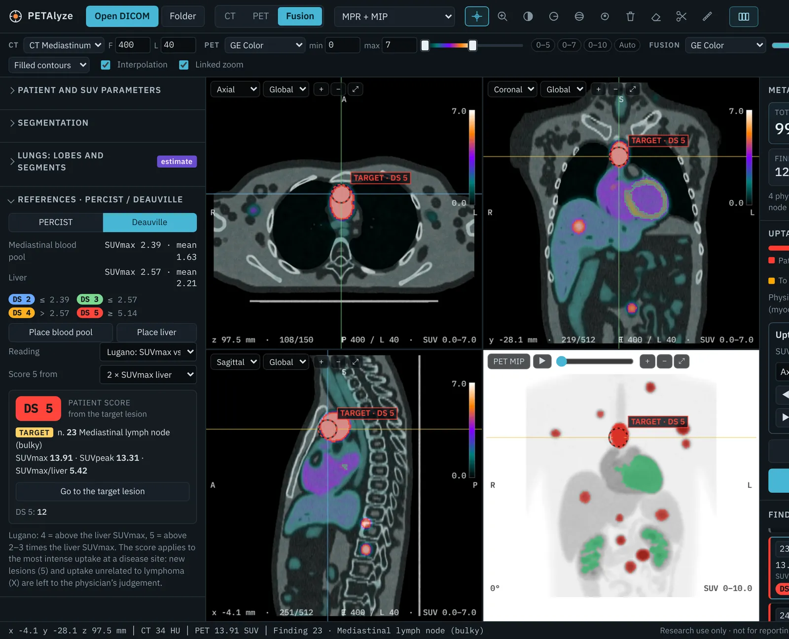
Place the liver and mediastinal blood pool references, or estimate the liver from the CT. Each lesion gets its 5-point score, and the most avid one is marked as the target lesion on the images, on the MIP and in the list.

Choose the Lugano reading (SUVmax against blood pool and liver) or qPET (SUVpeak over liver SUVmean).

Deauville panel with patient score DS 5 from the target lesion, and the target lesion marked on the axial, coronal and sagittal views.
Patient score from the target lesion, the bulky mediastinal node.

Report

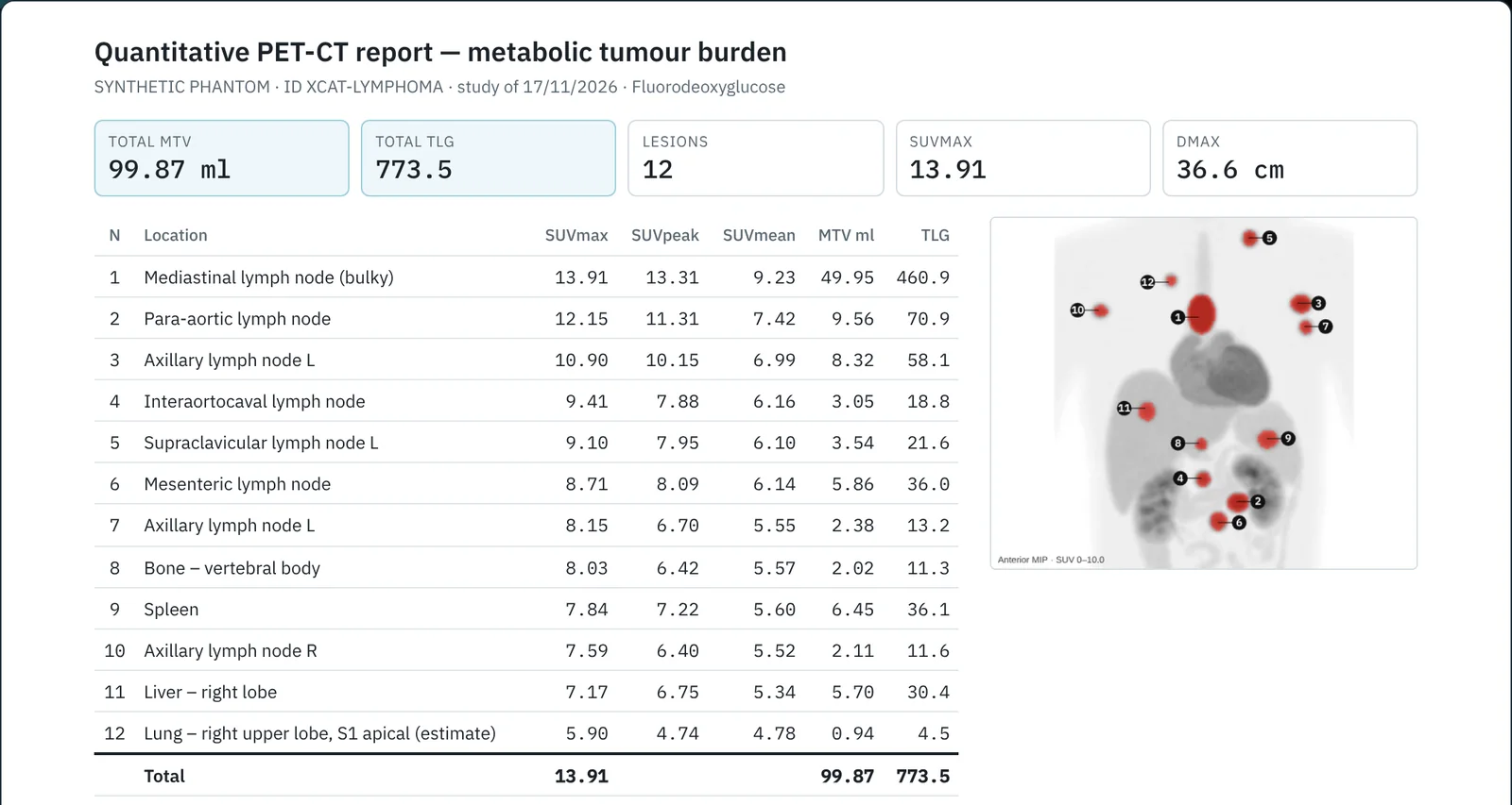
The MTV report lists the pathological lesions from the highest SUVmax, with the numbered MIP, the equivocal findings apart, the Deauville score and the segmentation method. Save it as HTML, or copy the text, a CSV table or a PNG of the views.

Quantitative PET/CT report with total MTV, TLG, lesion count, SUVmax and Dmax, a table of twelve lesions and the MIP with the lesions numbered.
The MTV report.

Lungs, lobes and segmentsexperimental

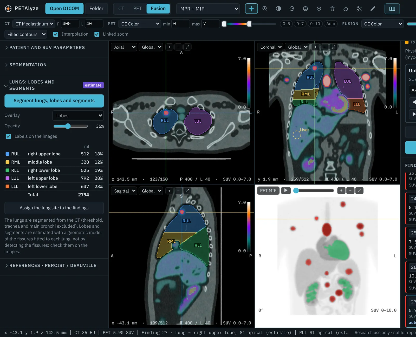
PETAlyze segments the lungs from the CT and estimates the five lobes and eighteen segments with a geometric model of the fissures, to name the site of lung findings. It is an estimate: check it on the images.

Lung lobes overlaid in colour on the axial, coronal and sagittal CT, with the lobe volumes listed.
Lobes overlaid on the CT, with their volumes.

Your images stay on your computer

This website delivers the program. Everything else happens in your browser.

This websitesends the PETAlyze page once
Your computerreads the DICOM files, processes them and saves what you export
  • DICOM files opened with Open DICOM or Folder, or dropped onto the window, are read directly from your disk and kept in the memory of the browser tab.
  • The code sends no data anywhere: nothing is uploaded, neither to this website nor to anyone else.
  • When you close or reload the tab, the images are cleared from memory.
  • PETAlyze stores only display preferences in your browser: colour scales, layout and segmentation method. No patient data.
  • Reports, CSV tables and PNG images go to the Downloads folder of whoever saves them. They contain the patient name read from the DICOM files, so they exist only on that computer.

Research use only

PETAlyze is intended for research purposes only. It is not a medical device and must not be used to report patient examinations or to make clinical decisions.

SUV and MTV values have been tested by comparing the results with those obtained on the same cases with syngo.via, but they are not validated for clinical use. Lung segmentation is experimental, and in general the software can make errors.

Users are responsible for ethics approvals, consent and data protection. We recommend working with anonymised or pseudonymised data.

Open a study in PETAlyze

Open PETAlyze